
Facial Surgical Intelligence

Understand the Structure.
Plan the Result.

Gain the vision others lack, and the advantage every surgeon wants. EVA 3D™ transforms not just how consultations are conducted, but provides tailored preparation for enhanced surgical outcomes.

Guided by trust, precision, and clear vision — built exclusively for board-certified facial plastic surgeons.
Apply for the Pilot Program

Request Your Consultation

Takes 60 seconds. Our concierge team follows up within one business day.

Thank you! Your submission has been received!
Oops! Something went wrong while submitting the form.
Facial Intelligence

How EVA 3D
Sees the Face

Six structural signals, mapped in real time — before a single incision.

EVA 3D™
Scan
Facial Volume Distribution
Asymmetry & Contour
Cheekbone Projection
Tear Trough Depth
Nasolabial Fold Structure
Temple Volume & Depth
Diagnostic Intelligence

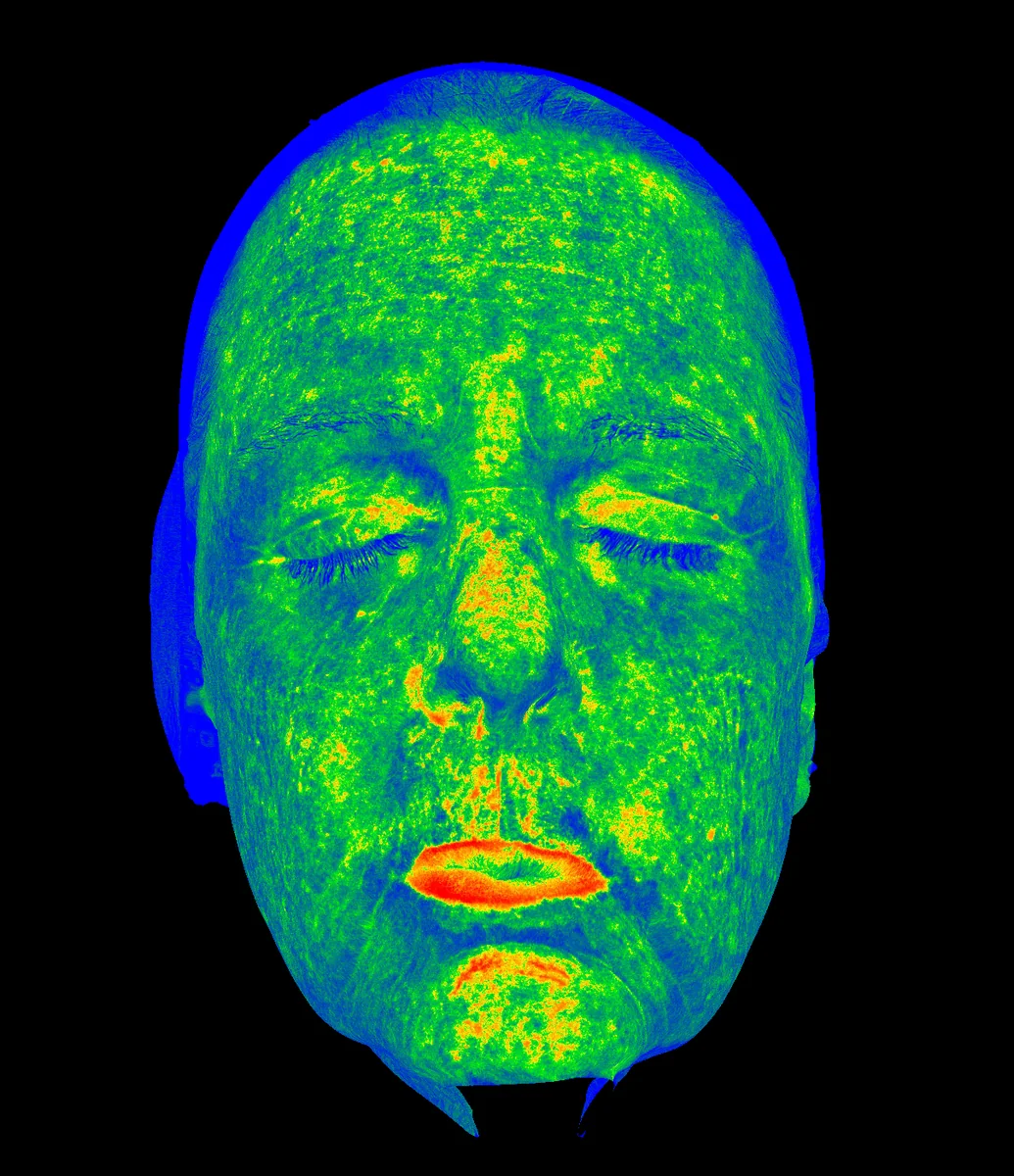
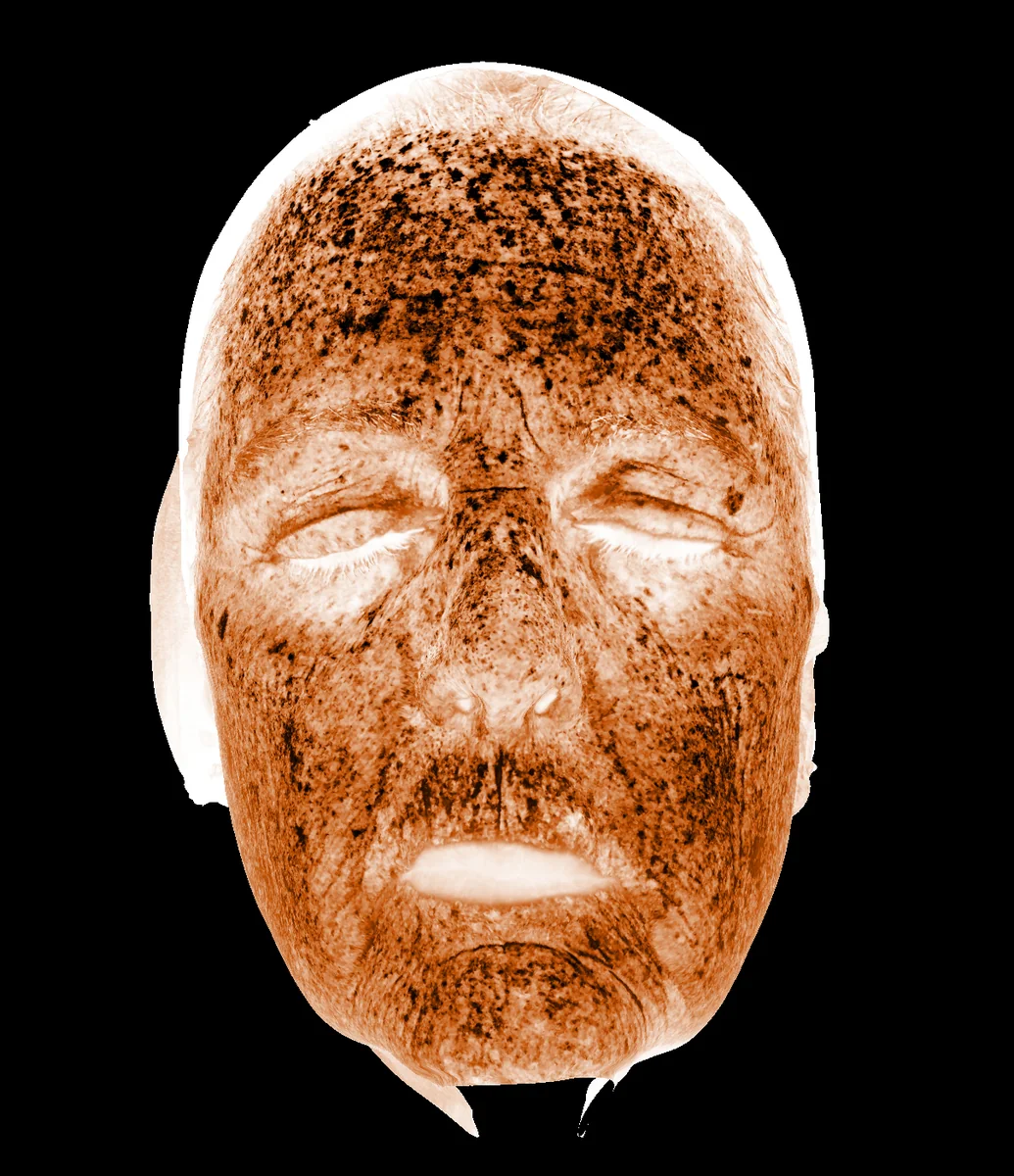
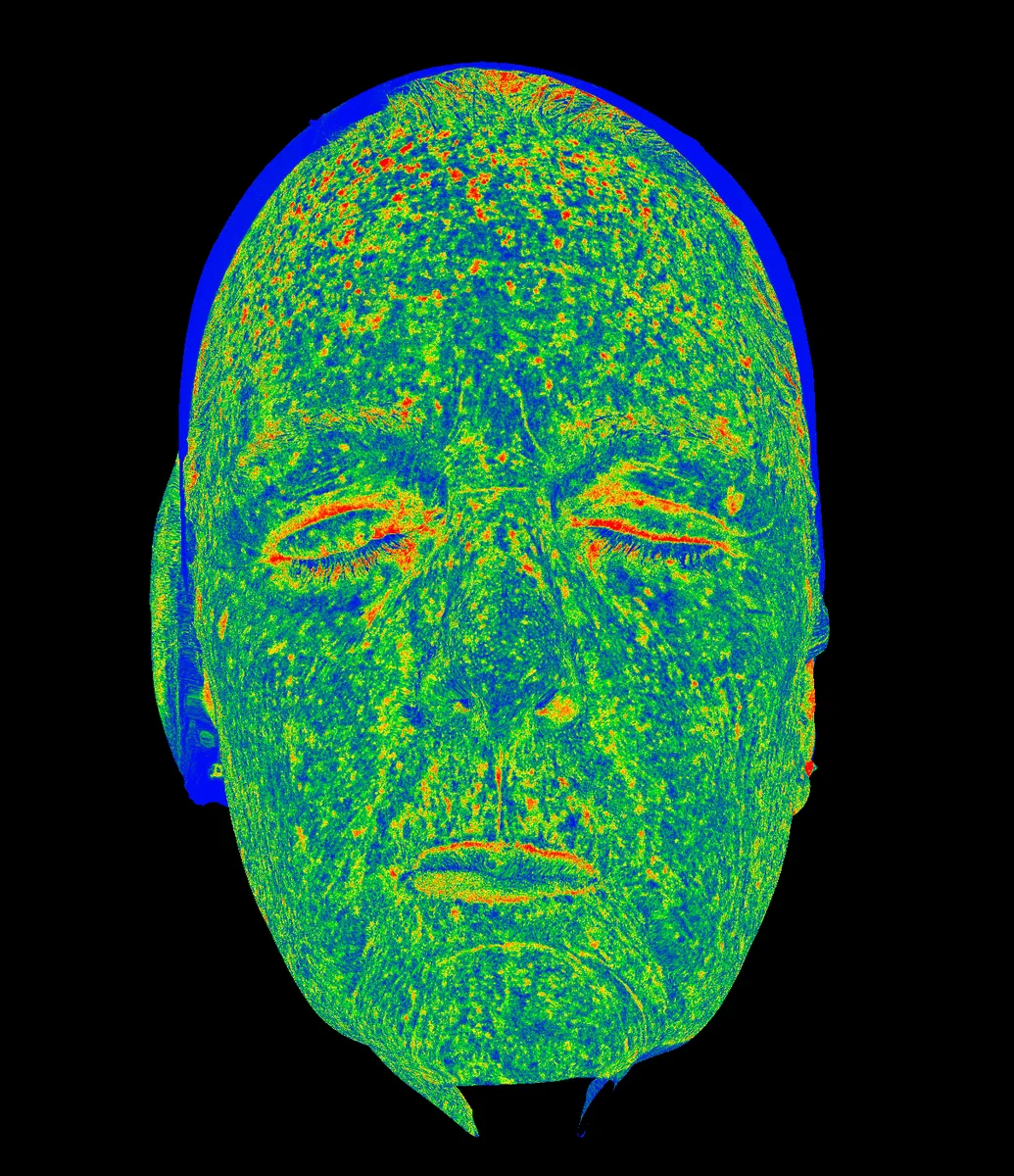
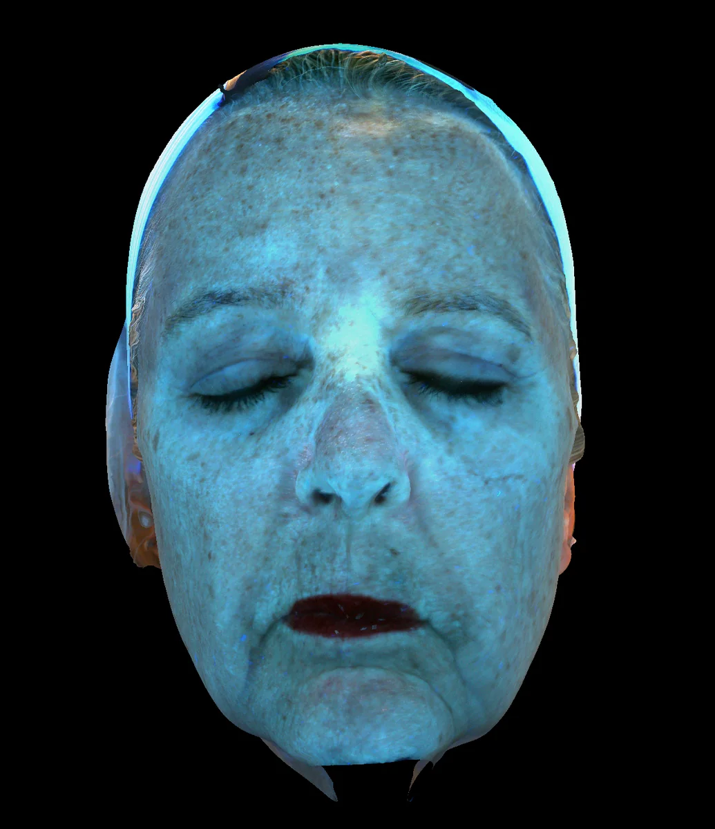
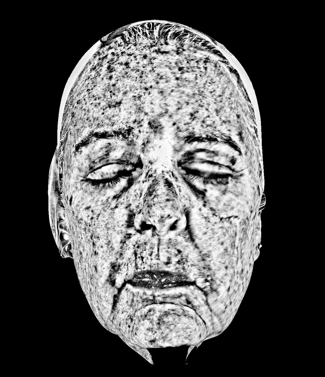
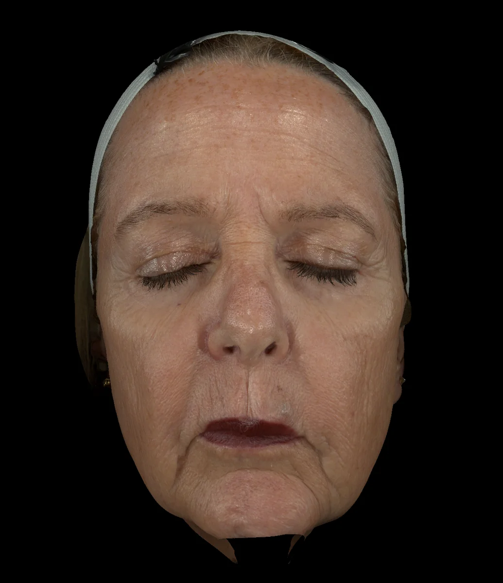
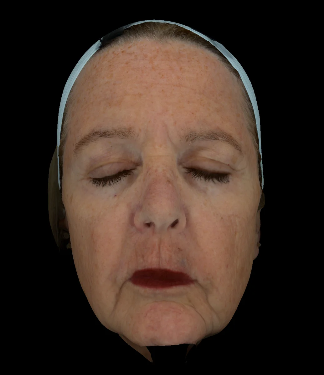
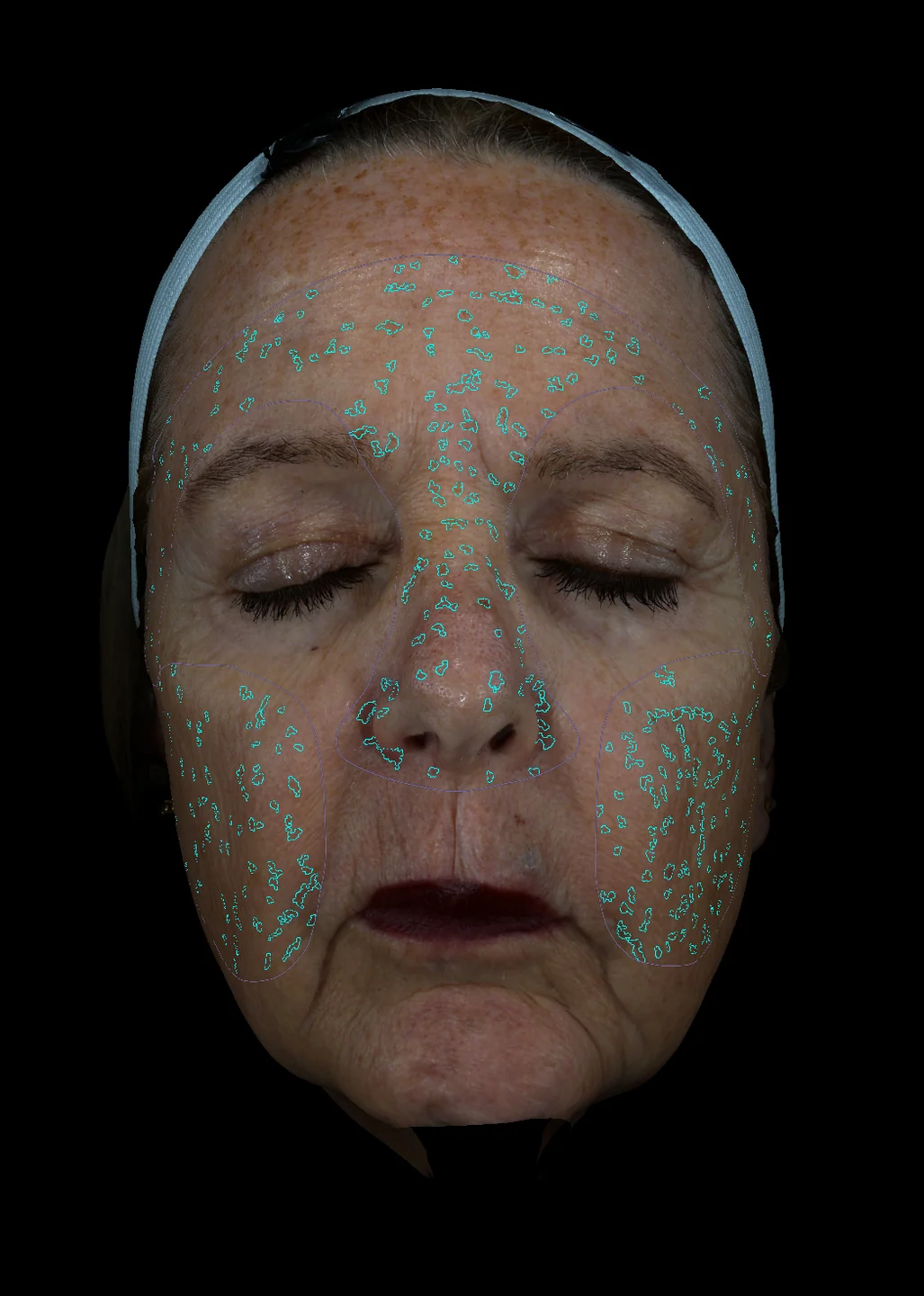
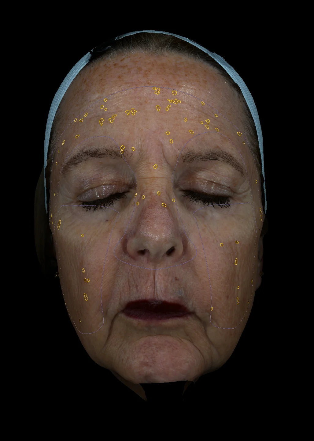
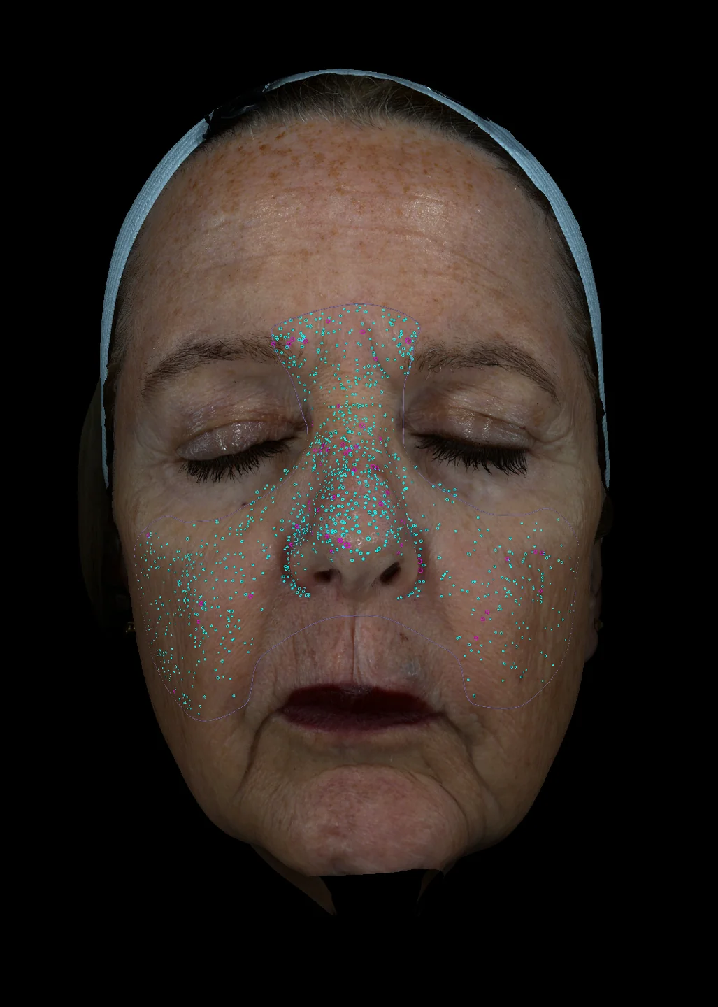
13 Light Spectrum Views. One Face.

Every EVA 3D scan captures the face across thirteen distinct light spectrums, each engineered to surface a different diagnostic layer, from surface texture to deep dermal chromophores. Tap a view to explore what it reveals.

Maximizes visibility of surface-level skin features.

Diagnostic Features
  • Pigmentation
  • Fine surface lines
  • Enlarged pores
  • Blackheads / comedones
  • Acne lesions
  • Redness and erythema
  • Sensitivity / irritation
  • Telangiectasia
  • Surface texture irregularities
  • Early dyschromia
  • Subtle surface roughness
Clinical Use
  • Highlights superficial color differences
  • Reveals fine textural changes
  • Ideal for baseline evaluation of surface-level abnormalities

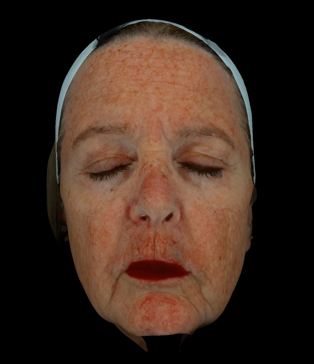
Replicates how the skin appears in normal daily environments.

Diagnostic Features
  • Pigment spots
  • Uneven skin tone
  • Fine lines and wrinkles
  • Enlarged pores
  • Sensitivity
  • Inflammation / erythema
  • Acne lesions
  • Telangiectasia
  • Blackheads / comedones
  • Visible texture irregularities
  • Overall surface-level disease burden
Clinical Use
  • Provides the most realistic, everyday appearance
  • Aligns patient concerns with objective clinical findings
  • Ideal for establishing a true-life baseline

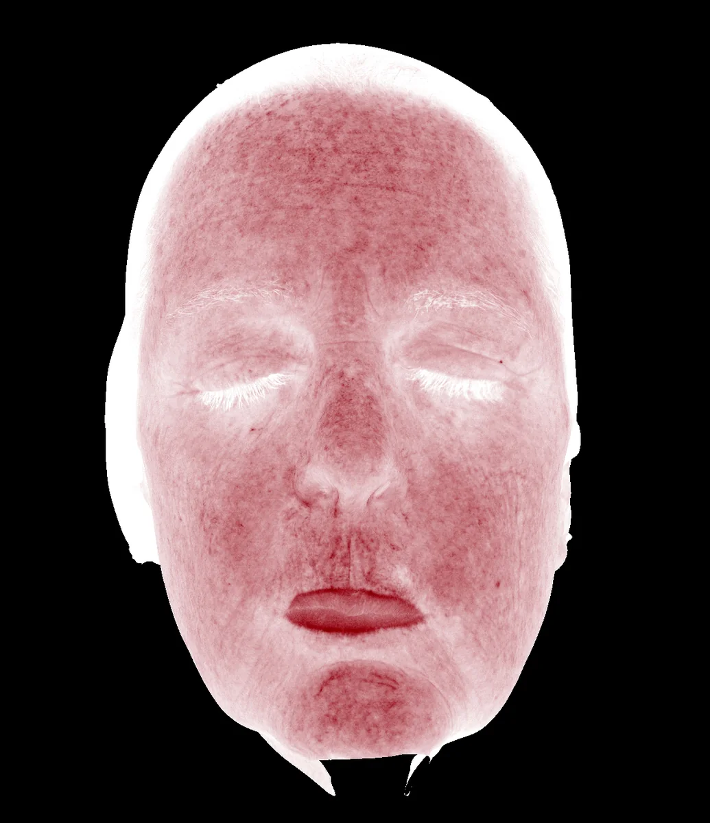
Highlights superficial hemoglobin distribution to reveal surface-level blood flow patterns.

Diagnostic Features
  • Superficial vascular dilation
  • Hemoglobin concentration patterns
  • Filamentous and reticular telangiectasia
  • Punctate vascular dilation
  • Rosacea-associated vascular patterns
  • Inflammatory vascular responses
  • Early erythema distribution
  • Surface-level redness networks
Clinical Use
  • Maps superficial hemoglobin patterns
  • Distinguishes diffuse redness from telangiectasia
  • Ideal for baseline vascular assessment and tracking changes over time

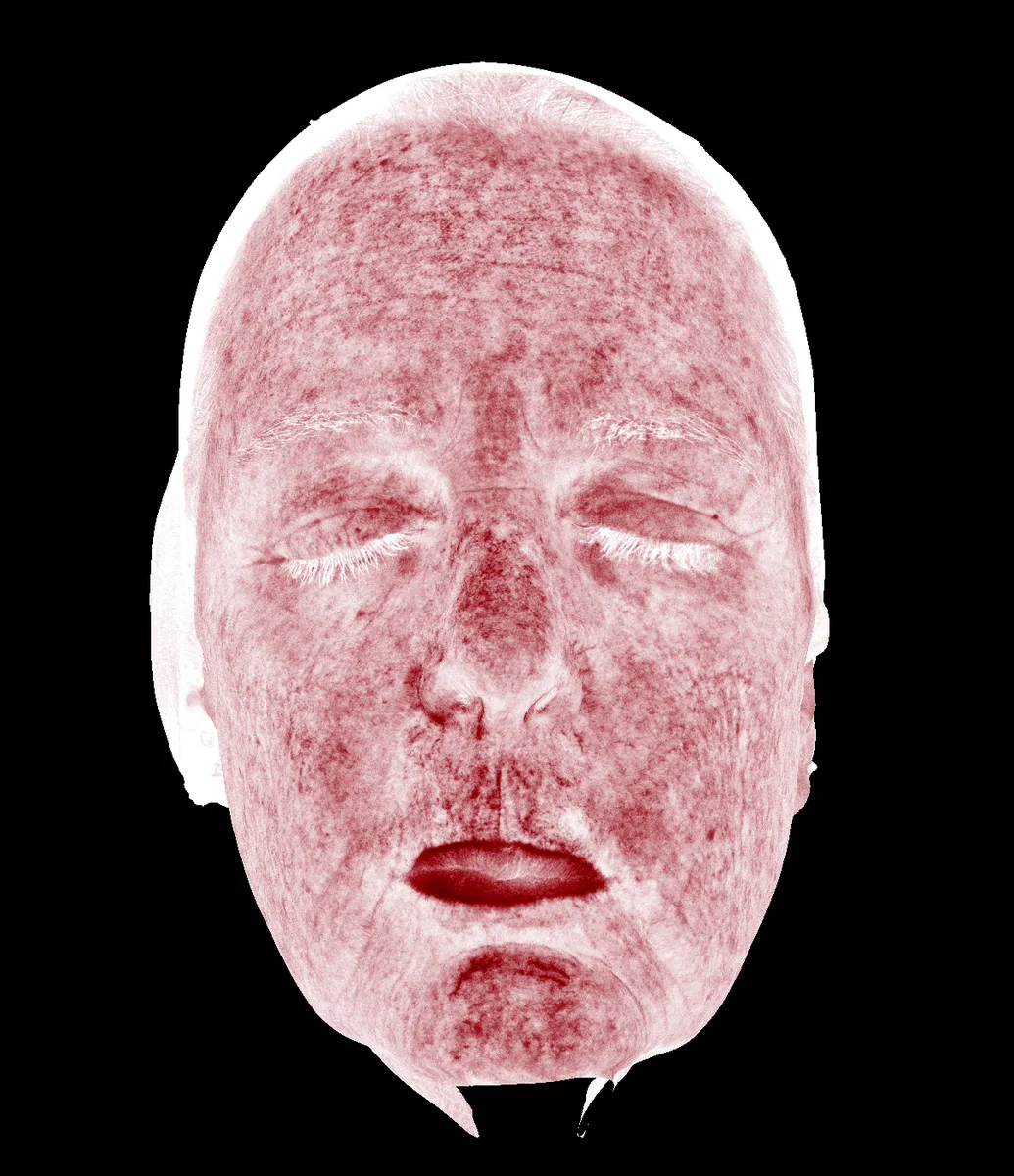
Visualizes hemoglobin distribution within the deeper dermal layers.

Diagnostic Features
  • Deep dermal hemoglobin concentration
  • Deeper inflammatory vascular patterns
  • Chronic or persistent redness
  • Subsurface vascular congestion
  • Sensitivity and inflammatory signatures not visible on the surface
  • Underlying vascular load contributing to rosacea
  • Hidden erythema beneath a clinically "mild" appearance
Clinical Use
  • Identifies deeper vascular congestion
  • Reveals chronic inflammation not visible on the surface
  • Useful when surface redness is minimal but deeper activity is suspected
  • Ideal for tracking persistent dermal erythema over time

Visualizes hemoglobin distribution in the upper dermal layers.

Diagnostic Features
  • Superficial (upper-dermal) vascular dilation
  • Early telangiectasia
  • Mild rosacea patterns
  • Sensitivity-related vascular responses
  • Barrier dysfunction indicators
  • Low-grade erythema
  • Early inflammatory changes
Clinical Use
  • Detects subtle or early erythema missed in standard lighting
  • Documents superficial inflammatory activity and sensitivity
  • Ideal for tracking short-interval treatment response in early rosacea or telangiectasia

Converts hemoglobin data into an easy-to-interpret heat scale.

Diagnostic Features
  • Color-graded inflammation severity
  • Blue = healthy baseline
  • Green = mild vascular activity
  • Yellow = moderate erythema
  • Red = severe inflammation
  • Overall redness distribution
  • Regional erythema hotspots
  • Rosacea flare zones
  • Comparative vascular intensity across areas
Clinical Use
  • Provides an instant visual severity index for erythema and rosacea
  • Enables comparison of inflammation across specific regions
  • Ideal for tracking changes and documenting treatment response over time

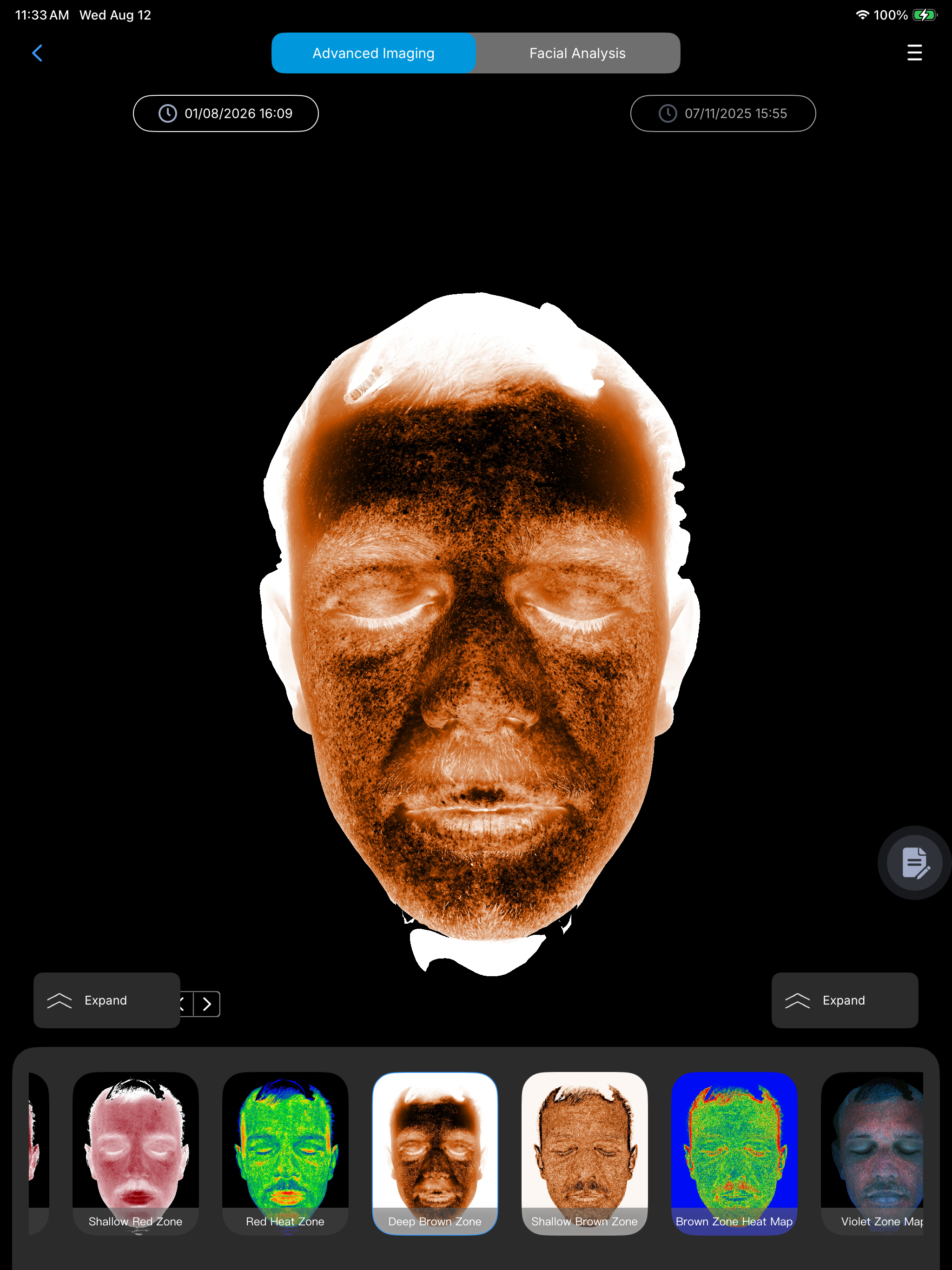
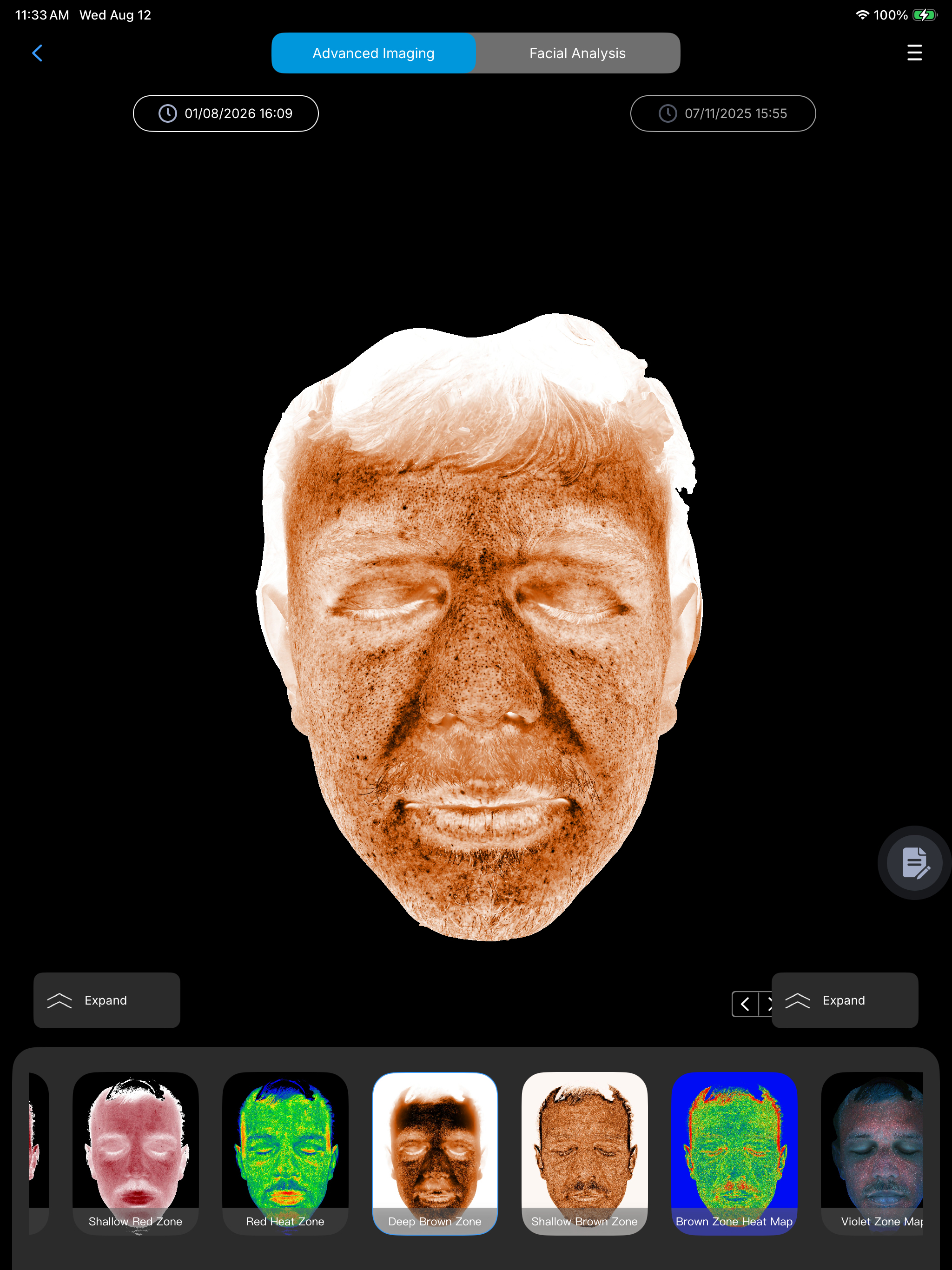
Enhances visibility of melanin located within the deeper dermal layers.

Diagnostic Features
  • Deep melanin concentration
  • Brown-black melanin deposits
  • Dermal hyperpigmentation
  • Localized deep pigment patches
  • Melasma (dermal or mixed type)
  • Deep post-inflammatory hyperpigmentation (PIH)
  • Pigmented nevi
  • Mixed-depth pigment patterns
  • Differentiation of deep vs. superficial dyschromia
Clinical Use
  • Distinguishes deep or mixed-depth pigment from superficial discoloration
  • Ideal for assessing melasma and deep PIH
  • Establishes a depth-weighted pigment baseline for long-term monitoring

Visualizes melanin concentrated in the epidermis.

Diagnostic Features
  • Superficial epidermal melanin
  • Early pigment aggregation
  • Freckles (ephelides)
  • Mild post-inflammatory hyperpigmentation (PIH)
  • Surface dullness
  • Uneven or blotchy skin tone
  • Early sun damage
  • Surface-level dyschromia patterns
Clinical Use
  • Captures early or surface-level pigment changes
  • Ideal for documenting epidermal dyschromia
  • Tracks tonal fluctuations between visits
  • Useful for identifying mild PIH and freckling patterns

Visualizes pigment severity and distribution across the skin.

Diagnostic Features
  • Color-graded melanin accumulation
  • Blue = healthy baseline
  • Green = mild pigment
  • Yellow = moderate pigment
  • Orange = high pigment
  • Red = severe melanin concentration
  • Spatial distribution of pigment load
  • Melasma severity zones
  • Hyperpigmentation density patterns
  • Early and advanced pigment clusters
Clinical Use
  • Provides an objective visual severity index for hyperpigmentation and melasma
  • Ideal for tracking pigment reduction or spread over time
  • Supports consistent, comparable visual or numeric trend monitoring

Reveals surface residues, microbial activity, and fluorescence-positive materials on the skin.

Diagnostic Features
  • UV fluorescence from product residue
  • Sunscreen residue
  • Microbial metabolites (e.g., Malassezia)
  • Porphyrins associated with acne bacteria
  • Oil buildup and clogged pores
  • Areas of poor cleansing or residual makeup
  • Fluorescent contrast between clean vs contaminated zones
  • Melanin and hemoglobin absorption (appear darker)
  • Subclinical pigment borders not visible in white light
Clinical Use
  • Identifies hygiene issues and product buildup
  • Detects microbial activity on the skin
  • Reveals subclinical dyschromia borders
  • Clarifies residue patterns that may interfere with diagnostics

Highlights areas where melanin, hemoglobin, and other UV-absorbing structures accumulate.

Diagnostic Features
  • Strong UV absorber regions (appear darker)
  • Melanin concentration zones
  • Hemoglobin-rich areas
  • Hidden or "occult" pigment foci
  • PIH boundaries
  • Melasma border definition
  • Nevus of Ota-type patterns
  • Vascular absorption clusters
  • Early sun damage
  • Areas of mixed pigment-vascular load
Clinical Use
  • Locates hidden pigment deposits not visible in standard light
  • Highlights vascular absorbers and UV-dense areas
  • Ideal as a corroborative map alongside brown/red modes
  • Helps define precise pigment boundaries and depth characteristics

Enhances specular (surface) reflection to highlight microtexture and contour irregularities.

Diagnostic Features
  • Surface wrinkles and rhytids
  • Fine lines
  • Enlarged pores
  • Acne scars (atrophic edges, texture breaks)
  • Surface oil and sebaceous shine
  • Microtexture irregularities
  • Surface roughness patterns
  • Contour discontinuities
Clinical Use
  • Accentuates surface texture by enhancing reflection
  • Ideal for evaluating wrinkles and fine lines
  • Highlights pore visibility and scar edges
  • Provides high-precision assessment of overall surface roughness

Suppresses surface glare to reveal deeper chromophore-based findings in the dermis and epidermis.

Diagnostic Features
  • Subsurface vascular activity
  • Skin sensitivity and inflammation
  • Telangiectasia
  • Acne activity under the surface
  • Barrier dysfunction indicators
  • Subsurface pigment spots
  • Melasma
  • Uneven skin tone
  • Dark circles (vascular/pigment)
  • Distinction between glare vs. true pathology
Clinical Use
  • Isolates subsurface chromophores (hemoglobin, melanin)
  • Reveals vascular and pigment pathology hidden by surface glare
  • Ideal for diagnosing erythema, rosacea, and telangiectasia
  • Excellent for identifying melasma and mixed-depth pigmentation
The Category Shift

Your Consultation Room Is Still in 2D.
Facial Surgery Isn’t.

Photos and mirrors ask patients to imagine the plan. EVA 3D™ makes your expertise visible, structured, and repeatable — before a single incision.

The 2D Consultation
  • Mismatched expectations
  • Difficulty earning trust
  • Elevated risk of complications
  • Longer consultation times
  • Lower conversion rates
The EVA 3D Consultation
  • Clearer communication through 3D modeling
  • Expectations aligned to anatomical limitations
  • Confidence built by addressing patient fears directly
  • Skin concerns identified before treatment
  • Higher conversion through visualized outcomes
The Platform

One System. Four Intelligent Modules.

EVA 3D™ is not a scanner. It is the complete facial surgical intelligence system — from first scan to final follow-up.

01

Diagnostic Studio

Evaluates skin health to identify visible, underlying, and predictive concerns that may need to be addressed before treatment.

02

Anatomy Studio

Focuses on facial anatomy and 3D modeling to simulate surgical outcomes for precise planning and realistic expectation setting.

03

Clinical Suite

Securely stores every generated case for side-by-side comparison, longitudinal review, and customized clinical reporting.

04

Intelligent Recovery

Tracks healing from Day 1 through Day 10+, turning post-operative care into a premium, high-touch extension of the consultation.

See it on your own patients — not a demo reel.

Apply for the Pilot Program

Designed for Facial Surgery

Purpose-built to support consultations for:

  • Facelift
  • Deep Plane Facelift
  • Neck Lift
  • Rhinoplasty
  • Fat Grafting
  • Injectables
  • Full Facial Rejuvenation

3D Visual Intelligence

EVA 3D™ reveals what matters most in facial structure:

  • Facial Volume Distribution
  • Asymmetry & Contour Balance
  • Cheekbone Projection
  • Tear Trough Depth
  • Nasolabial Fold Structure
  • Temple Volume & Depth
0.1mmScan Precision
0.005mlVolume Analysis Accuracy
7-StepStructured Consultations
2-DayTeam Certification Program
Trusted Faculty

The Clinical Vanguard

EVA 3D™ is used and taught by leading facial plastic surgeons across the country.
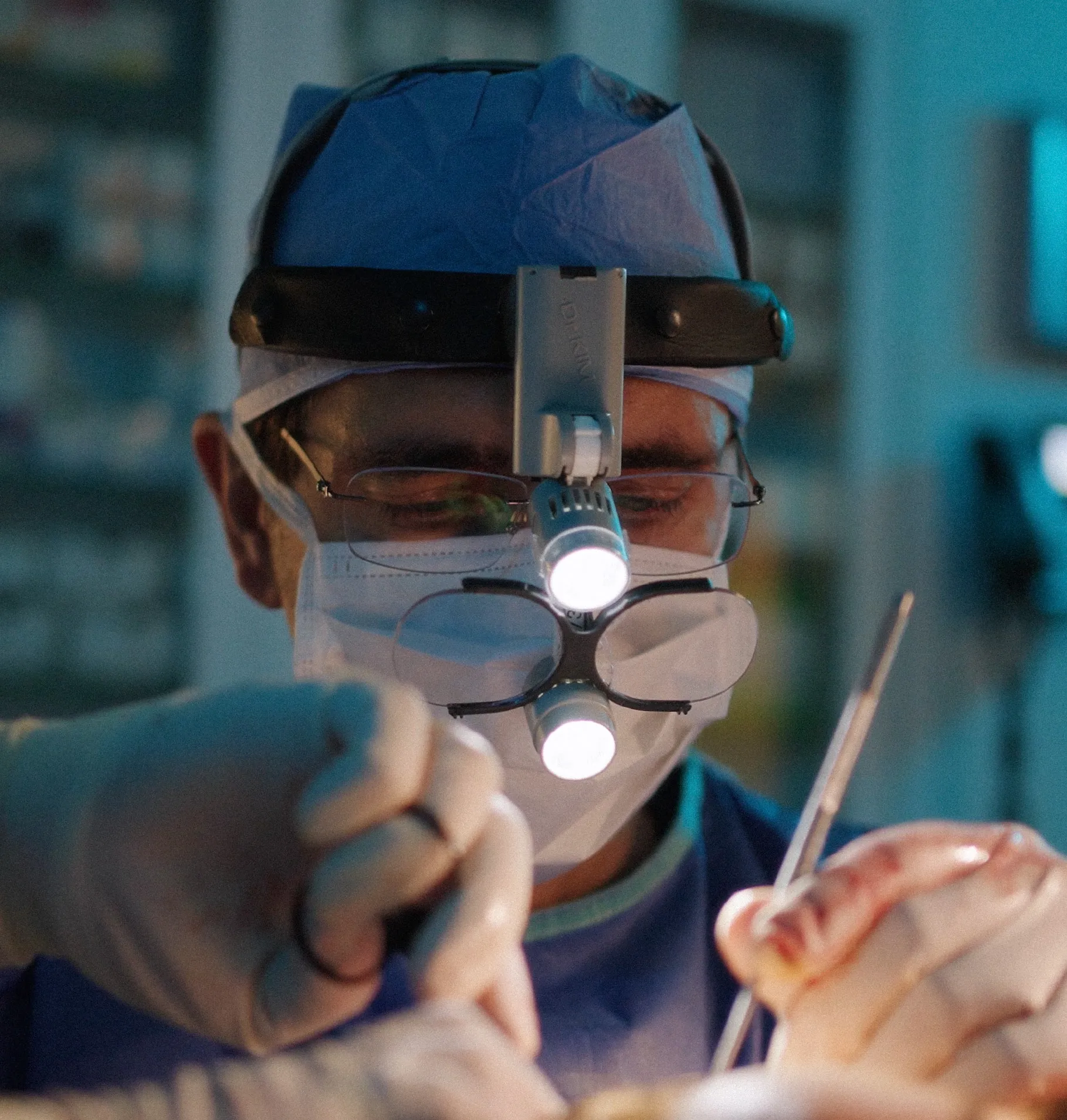
Dr. Ramtin Kassir

Ramtin Kassir, MD, FACS

Clinical VanguardNew York City, USA
Dr. Steven Camp

Steven Camp, MD, FACS

Clinical VanguardTexas, USA
Dr. Rod J. Rohrich

Rod J. Rohrich, MD, FACS

Clinical VanguardTexas, USA
Dr. Richard Balikian

Richard Balikian, MD, FACS

EVA University FacultySan Diego, USA
Dr. Kevin Sadati

Kevin Sadati, DO, FACS

Clinical VanguardCalifornia, USA
Dr. Kristy Hamilton

Kristy Hamilton, MD, FACS

Clinical VanguardTexas, USA

Join the surgeons already
teaching with EVA 3D™

Apply for the Pilot Program
Education & Certification

EVA University

Every practice completes a structured two-day certification — hands-on Anatomy Lab training and cadaver dissection, run in partnership with the NYC Facelift & Rhinoplasty Master Course and NYU Langone Health.

Explore Certification

“Understand the Structure.
Plan the Result.”

The EVA 3D Standard

Trusted by Top Surgeons.
Now It’s Your Turn.

Apply for the EVA 3D Facial Surgery Pilot Program and bring intelligent consultation planning to your practice.

Request a Demo

Facial surgical intelligence

See What
Others Can’t.

EVA 3D gives facial plastic surgeons an objective three-dimensional view of facial structure, skin health, and change over time—so patients can understand the plan before treatment and see the truth after it.

Understand the Structure. Plan the Result.

Patient in a clinical consultation setting
Objective view · Structure made visible

The consultation gap

Your patient is three-dimensional.
Your consultation should be too.

Traditional vs EVA 3D™ What appears subtle in a photograph can become measurable in three dimensions.
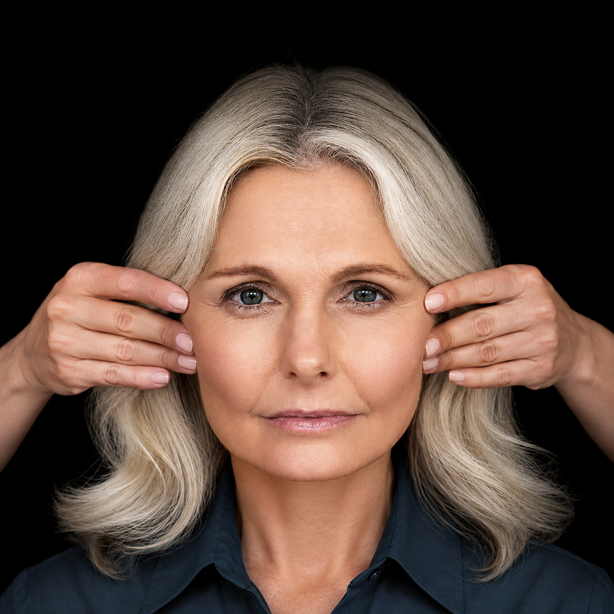
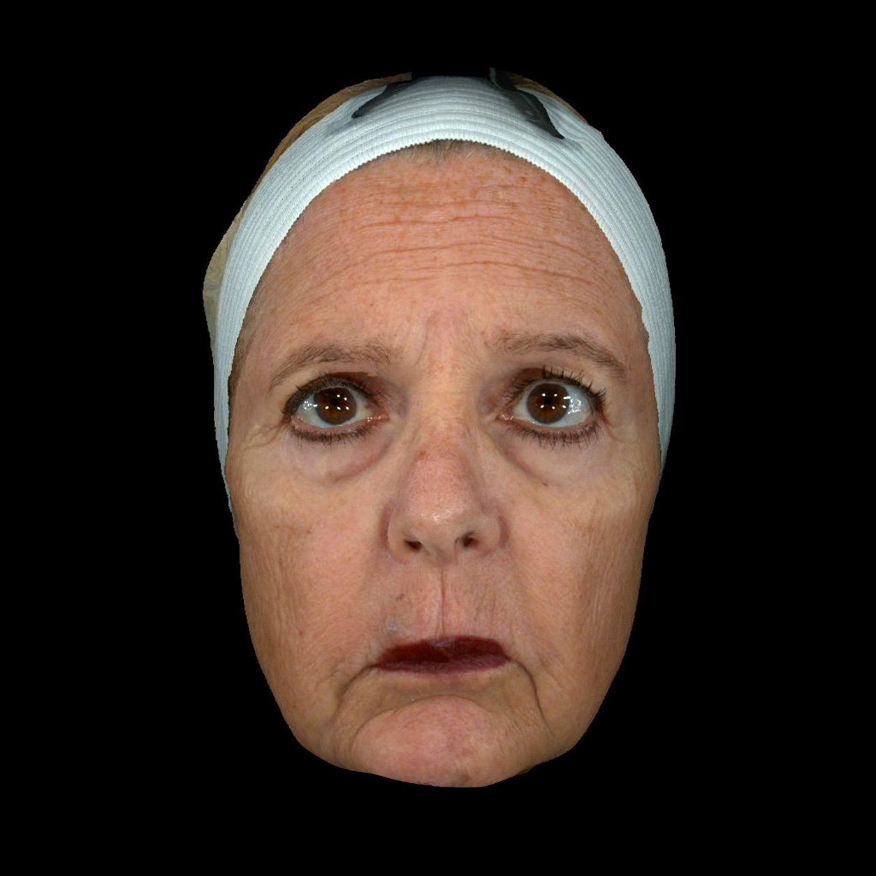
Traditional two-dimensional patient photograph

A traditional consultation relies on observation, explanation, and the patient’s imagination.Without objective measurements, subtle asymmetries and structural differences can be difficult to communicate clearly.
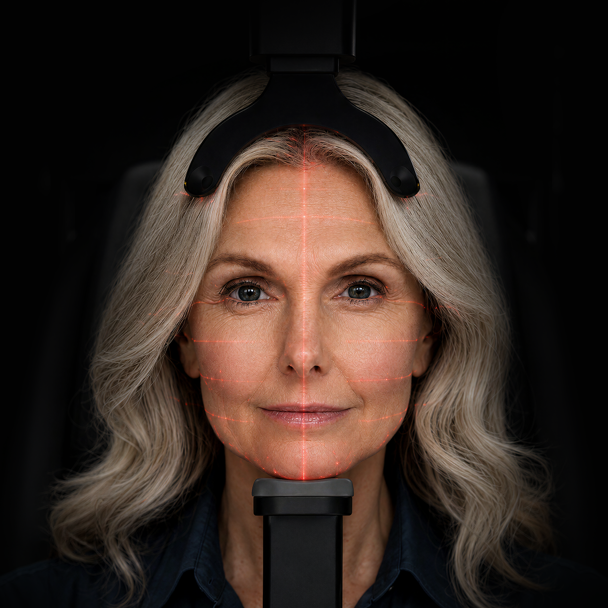
Patient view with an illustrative structural analysis overlay
Symmetry
Volume
Structure

EVA 3D turns what the surgeon sees into measurable, visual insight.Analyze symmetry, structure, volume, proportions, and more to create a more precise consultation, align expectations, and support better treatment planning.

The EVA 3D platform

Take a closer look.

One platform. Multiple ways to understand the face.

Diagnostic Studio™

Understand what the surface can reveal.

Each light-spectrum view helps the surgeon discuss pigmentation, vascular redness, inflammation, wrinkles, sensitivity, and pores.

Clinical use

Red Blood

  • Maps superficial hemoglobin patterns.
  • Distinguishes diffuse redness from telangiectasia.
  • Ideal for baseline vascular assessment and tracking changes over time.
Different wavelengths reveal different layers of skin health.

Anatomy Studio™

Move from structural analysis to surgical design without leaving the consultation.

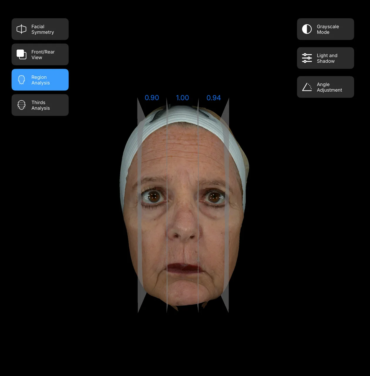
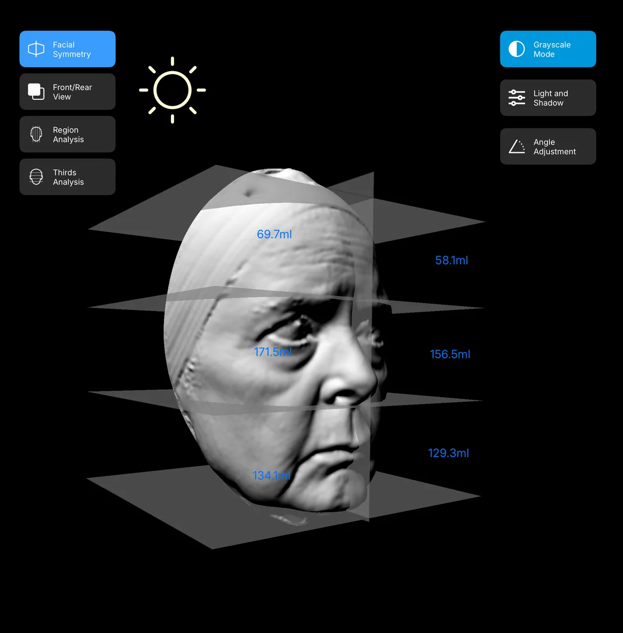
Facial Structural Analysis

See the face as a surgical map.

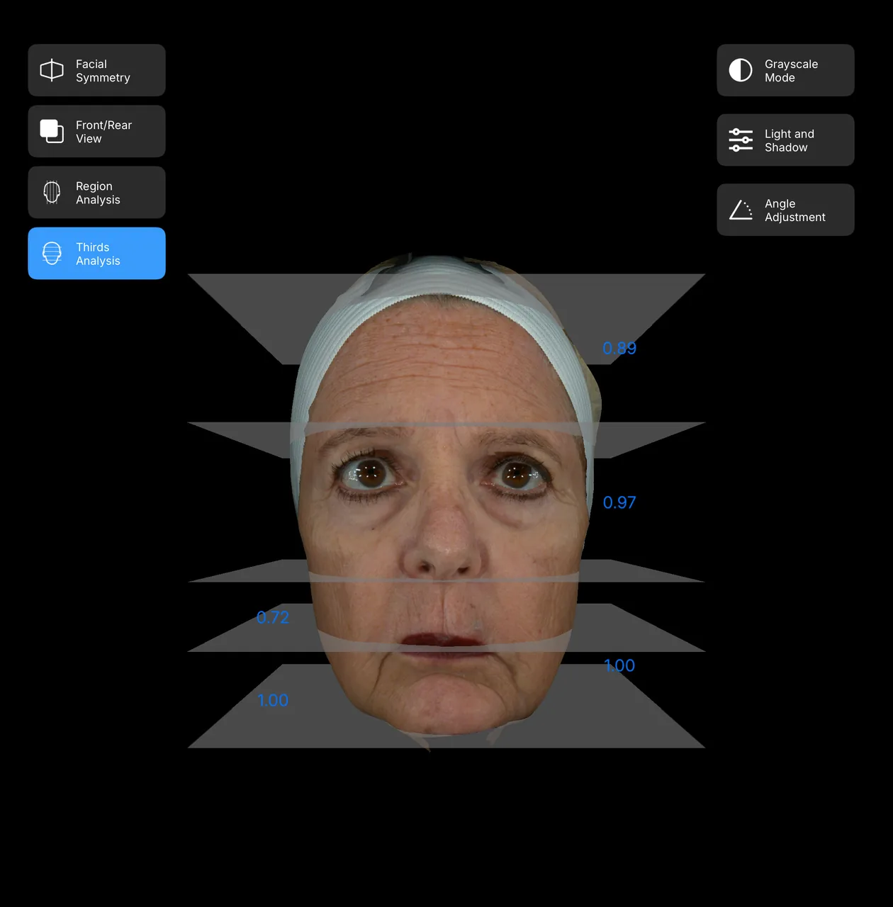
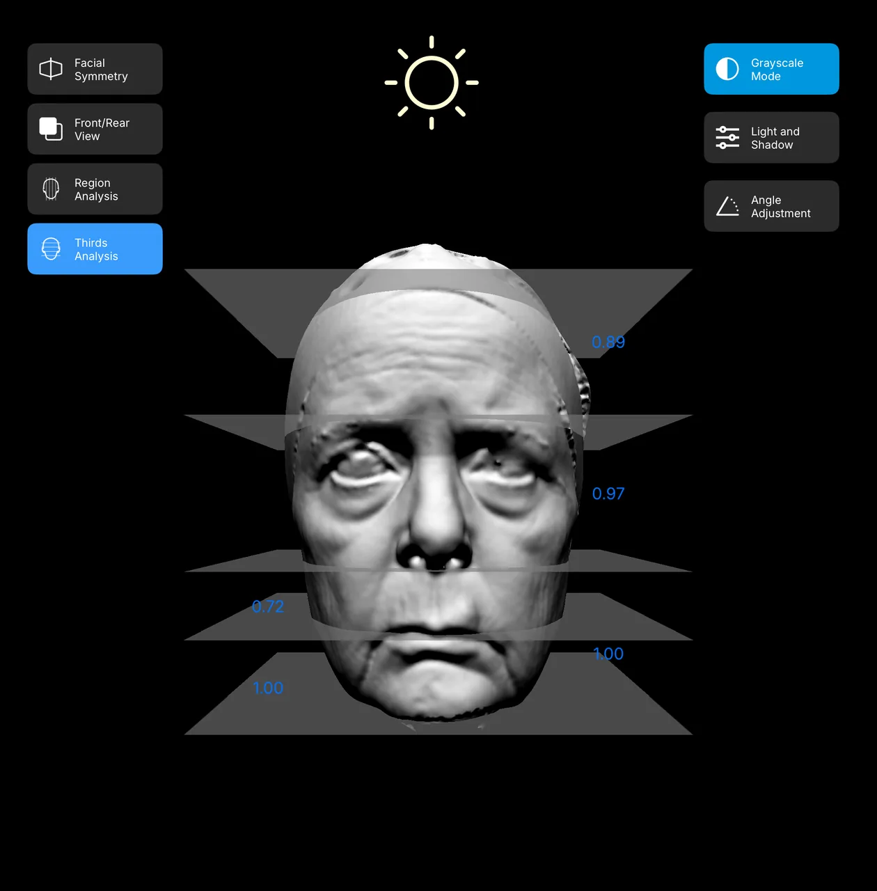
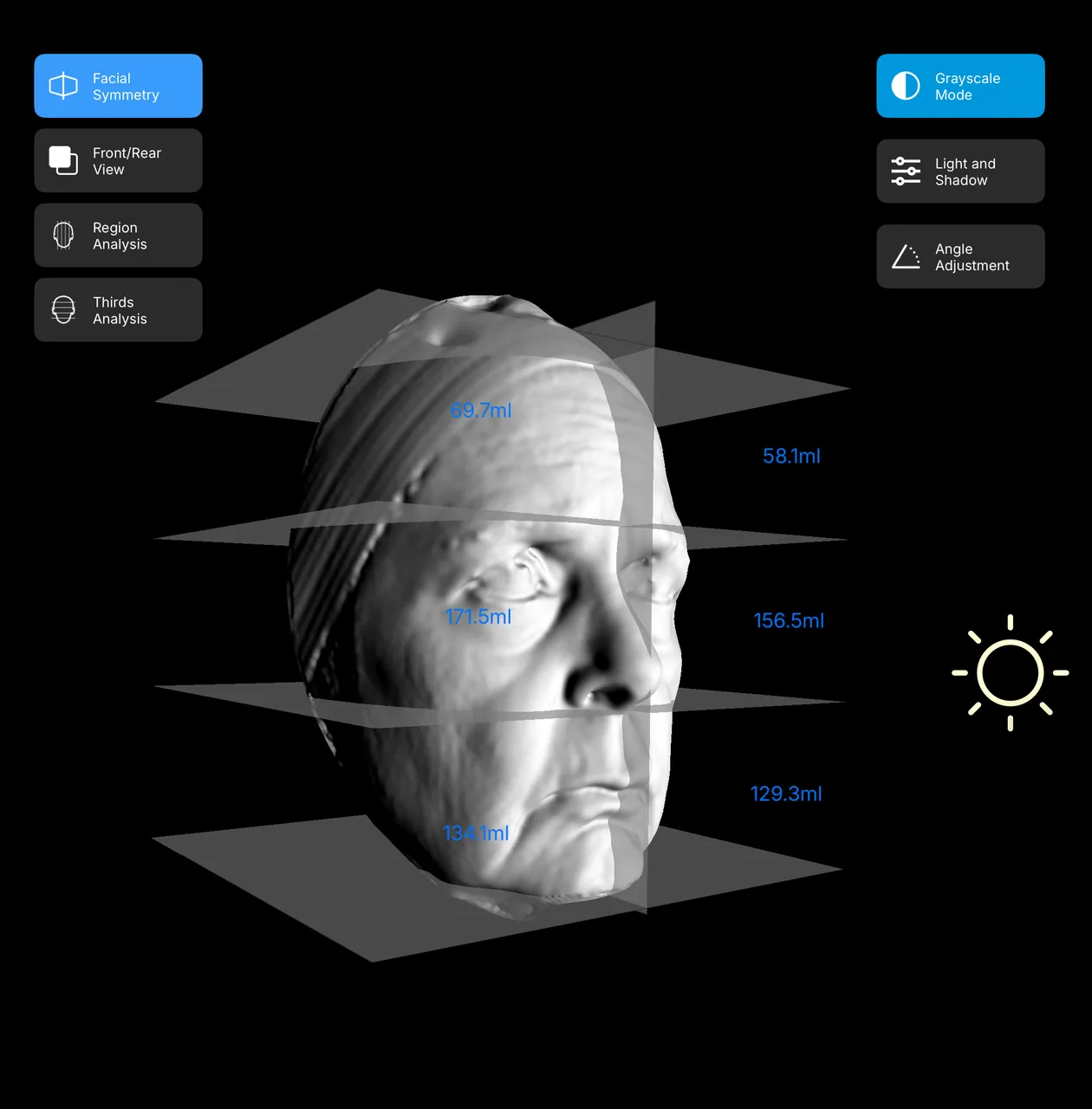
Before I recommend a procedure, I need to understand the face in three dimensions—not as a single photograph. EVA 3D separates proportion, surface topography, and volume so I can explain the anatomy behind my recommendation with the patient beside me.

01
Proportion, made visible

Regional and facial-thirds analysis reveals relationships between the upper, middle, and lower face, helping me evaluate asymmetry while keeping the whole face in view.

02
Surface, without distraction

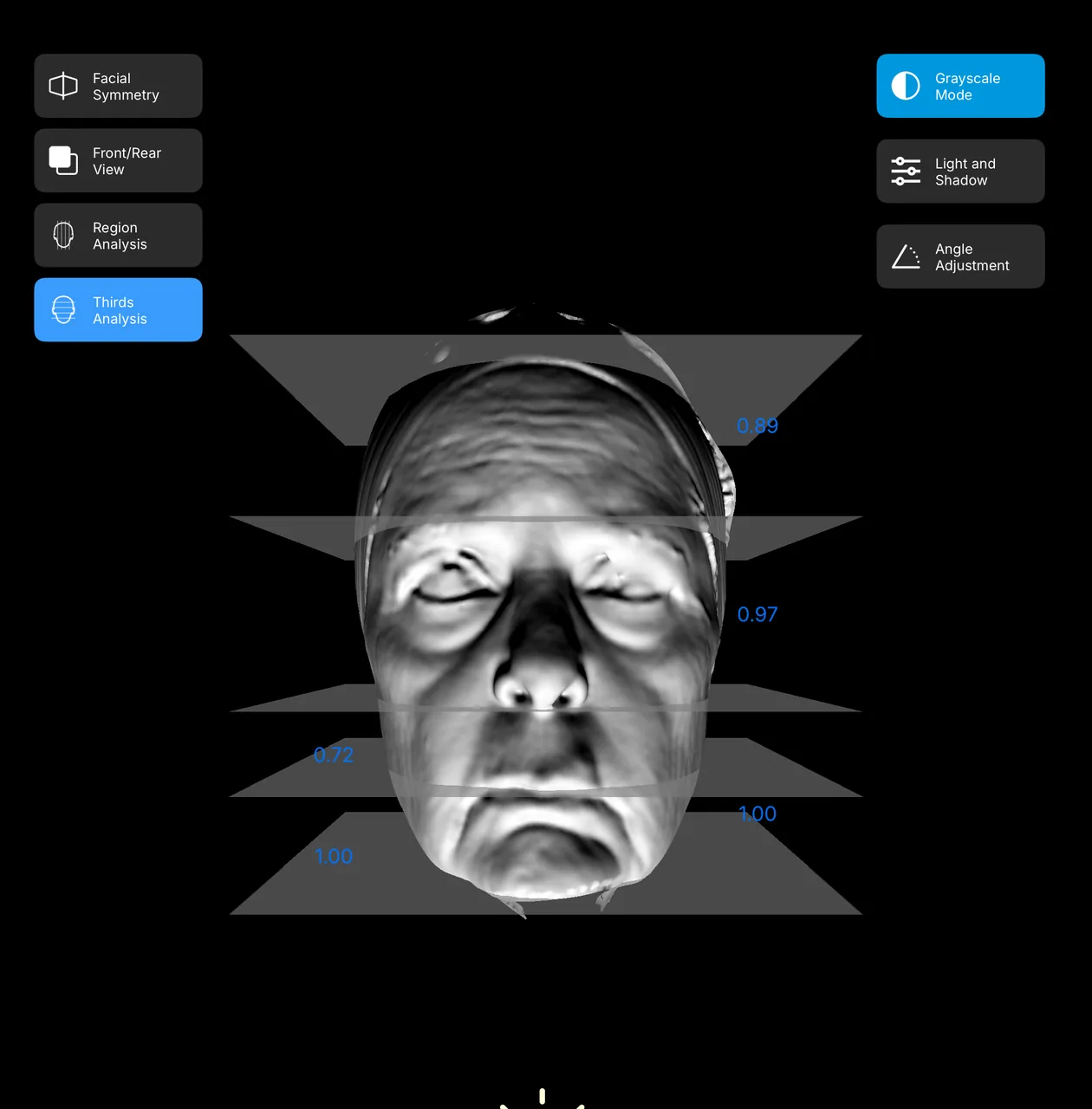
Grayscale removes color and lets me review the 3D surface under changing light. Moving the light source exposes contour irregularities, shadow transitions, and areas that deserve closer surgical consideration.

03
Precision by the millimeter

Linear and volumetric measurements turn an impression into a reproducible baseline. I can quantify the regions we are discussing, compare sides, and document the plan with millimeter-level precision.

“The consultation becomes more specific: the patient can see what I see, and I can explain why it matters.”
Aesthetic design Proposed Rhinoplasty Change
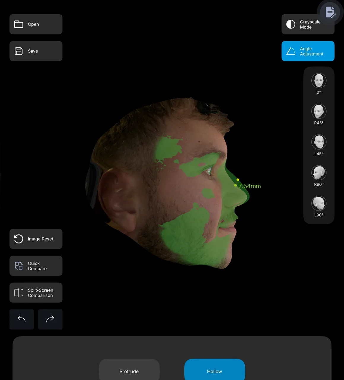
Male profile with green rhinoplasty design overlay and proposed 7.54 millimeter bridge change
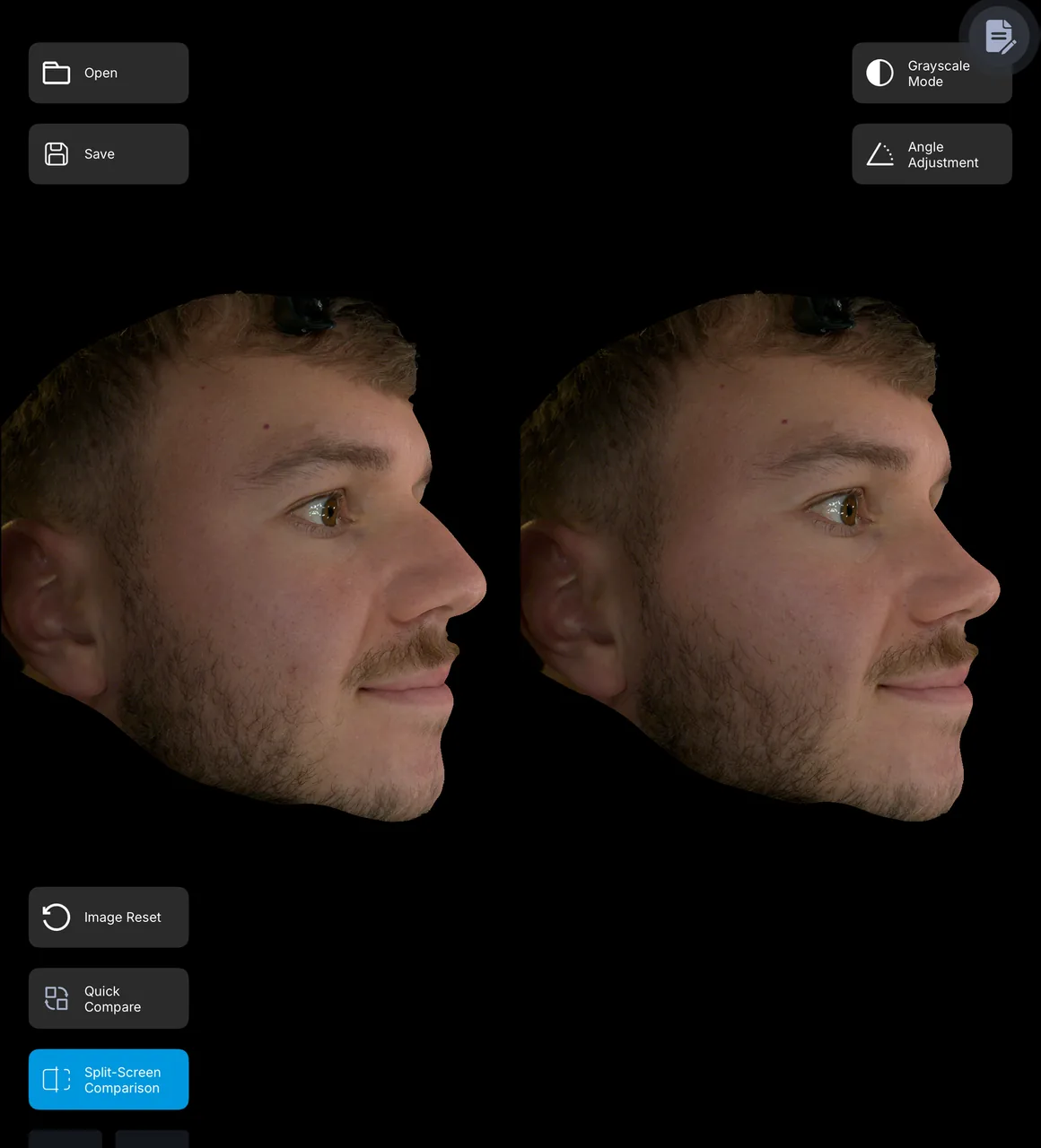
Male baseline and designed profile shown side by side

Aesthetic Design

Show the plan before the procedure.

In a rhinoplasty consultation, I can design the intended contour directly on the patient’s three-dimensional image, then review the proposed change from the same angles I use in surgical planning.

7.54 mm

The green overlay identifies the regions affected by the design. In this example, the measurement records a proposed 7.54 mm reduction from the superior nasal bridge—making the change concrete and reviewable rather than subjective.

01
Align expectations

Baseline and designed views sit side by side so the patient and I can agree on direction, proportion, and restraint before surgery.

02
Document the rationale

Measurements and affected zones create a clear visual record of what was discussed and why the proposed change fits the face.

03
Strengthen the consultation

When patients understand both the anatomy and the intended change, they ask better questions and can decide with greater confidence.

“EVA 3D turns my surgical judgment into a consultation the patient can see, understand, and remember.”

Design visualization supports communication and planning; it does not guarantee a surgical outcome.

EVA MARK™

One visual workspace for surgeon-directed planning and automated facial analysis.
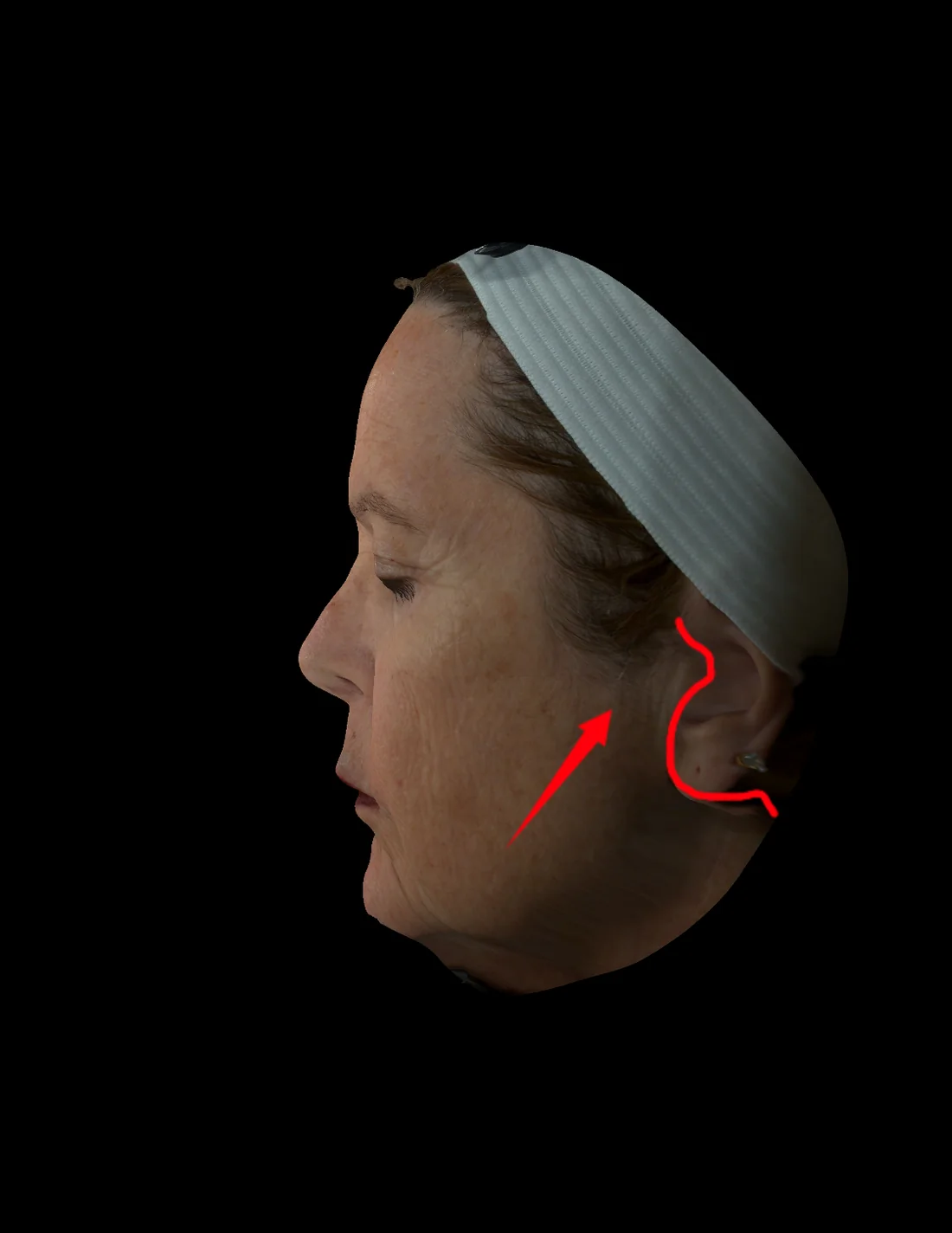
SURGEON-DIRECTED PLANPhysical marking, translated to 3D
● Editable annotation
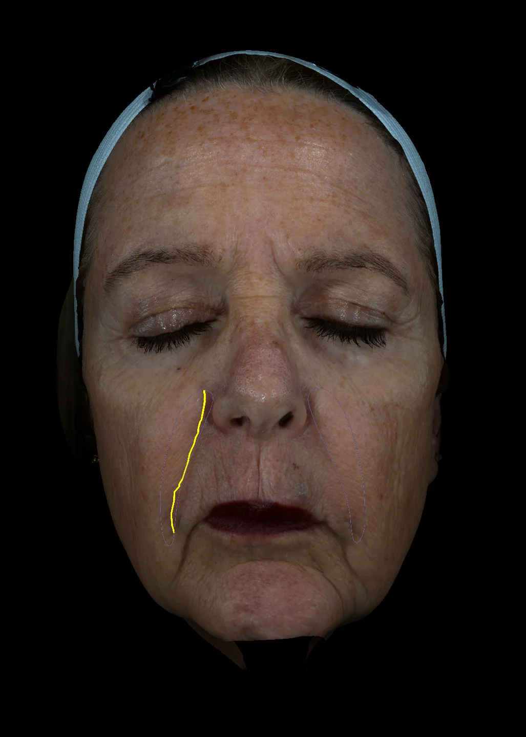
EVA Mark surgeon-drawn lift vector and periauricular boundary on a three-dimensional patient profile
Draw · rotate · explain
01Incision path
02Lift vector
03Boundary
04Clinical note
SURGEON MARKING

Show the patient exactly where the plan lives.

I can recreate the physical surgical marking plan on the patient’s three-dimensional image—then rotate the face and explain the incision, vector, boundary, and operative landmarks from the angle that matters.

01
Translate judgment into a visual plan

Connect the marks I make on the patient with the anatomy we are discussing.

02
Annotate from surgical angles

Add freehand lines, arrows, shapes, and labels directly to the 3D record.

03
Make consent conversations clearer

Give the patient a precise visual reference they can understand and remember.

“Instead of asking the patient to imagine my plan, I can point to it.”

EVA Mark supports consultation, annotation, and planning communication; it is not surgical navigation.

Deep Brown Zone facial imaging after laser treatment showing reduced visible sun damage
Deep Brown Zone facial imaging before laser treatment showing visible sun damage
BeforeAfter
Before 50% · After 50%

Laser Treatment Comparison

See less sun damage.

Drag across matched Deep Brown Zone images to compare the skin before and after laser treatment. The after view shows fewer concentrated areas of visible pigmentation associated with sun damage.

Make treatment progress visible.
Patient record · Longitudinal view
  1. ConsultationDiagnostic + Anatomy
  2. Pre-opEVA Mark plan
  3. Post-opRecovery scans
  4. ResultsObjective comparison
AFTER-VISIT REPORT

Your progress, available after every visit.

Patients receive a private diagnosis report after each scan to review findings, compare visits, and follow changes over time.

Open full report ↗

Clinical Suite™

Every patient. Every scan. One clinical record.

Organize scans, diagnostic findings, measurements, markings, and comparison records into a longitudinal view of the patient journey.

Clinical context, connected over time.

Intelligent Recovery™

Follow healing instead of guessing at it.

Track pre-op through post-op scans across the clinical recovery schedule, making progress visible and easier to discuss.

Recovery is measurable. Progress becomes visible.

The intelligent consultation

From scan
to surgical understanding.

013d facial analysis machine EVA 3D

Capture

Clinic staff completes the scan before the surgeon enters.

The surgeon arrives as the analyst, not the technician.
023D  facial analysis diagnostic studio

Diagnose

Diagnostic Studio reveals skin-health information across thirteen light-spectrum views.

033D  facial analysis EVA University

Understand

Eva University is a full proof guide to understanding how EVA3D can deliver better consultations.

043D  facial analysis anatomy studio

Plan

Anatomy Studio allows you to mark and explains the approach directly on the patient’s scan.

053D  facial analysis clinical suite

Track

Results and recovery become part of the patient’s longitudinal clinical record.

Objective comparison

Before. After.
And everything between.

Move beyond side-by-side photography.

SURGEON PERSPECTIVES

See EVA 3D™ through a surgeon’s eyes.

Choose a surgeon below to hear how EVA 3D supports clearer consultations, planning, and measurable outcomes.

Surgeons in their own words
Dr. Richard Balikian
“Surgery is not just about technique—it’s about pre-planning.”

Richard Balikian, MD, FACS

Video coming soon

Dr. Steven Camp
“Real-time data helps us understand treatment progress and deliver better results.”

Steven Camp, MD

Video coming soon
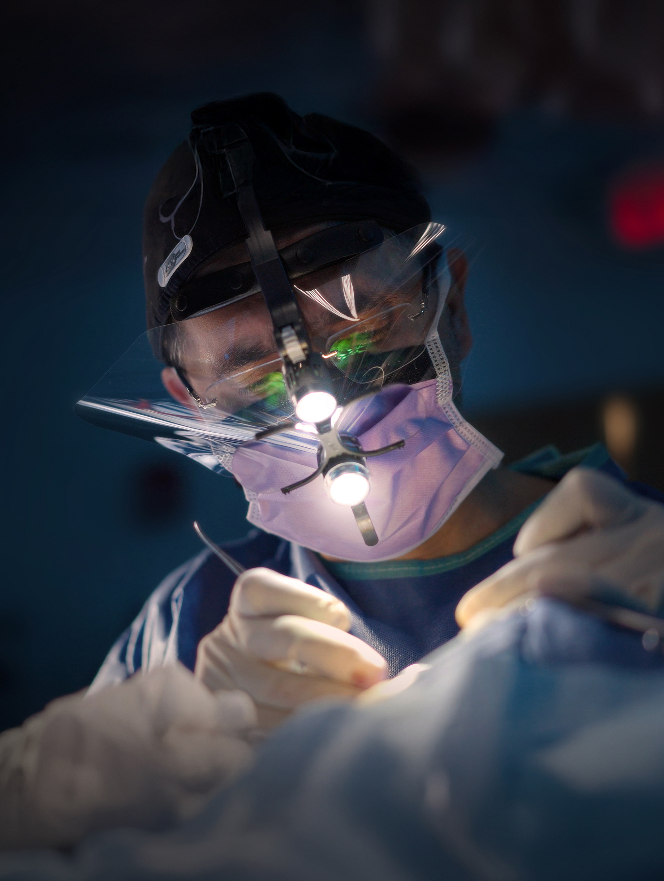
Dr. Ramtin Kassir
“I saw a 50% increase in patient conversions after implementing EVA3D™ into my consultations.”

Ramtin Kassir, MD, FACS

Video coming soon

Patient confidence

Patients trust
what they can understand.

Instead of asking a patient to imagine what the surgeon sees, EVA 3D allows the surgeon to show the anatomy, measurements, and findings directly.

01

See

Make invisible information visible.

02

Understand

Translate clinical judgment into something the patient can follow.

03

Decide

Create clearer alignment before treatment.

The system
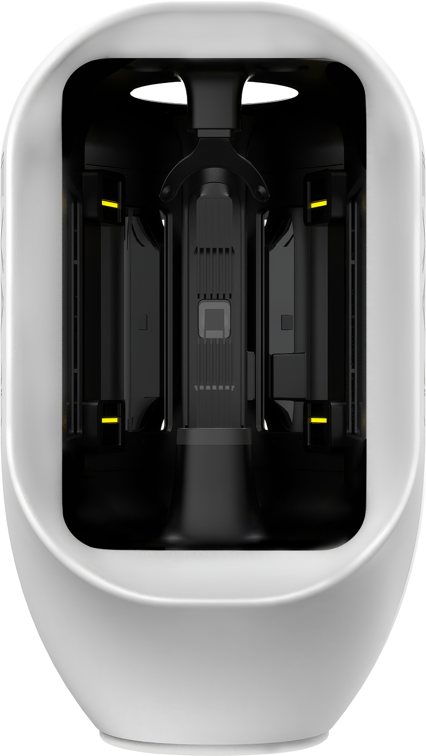
Precision deserves
the right instrument.

EVA 3D Facial analysis device for plastic surgeons consultations
Controlled captureConsistent imaging conditions
32-inch displayClinical detail at consultation scale
iPad + penPlan and explain in real time

The booth, display, table, processor, iPad, and pen are designed as one consultation environment.

Facial imaging system
Controlled, repeatable image capture.
Height-adjustable table
Patient comfort and procedural consistency.
Adjustable monitor arm
Position clinical detail where surgeon and patient can review it together.
THE HARDWARE01 / 03
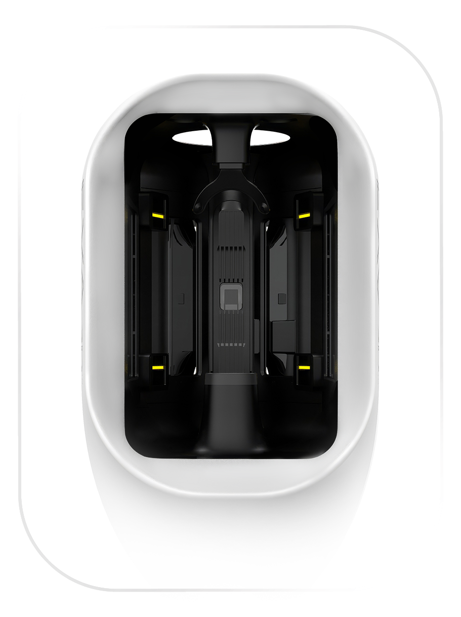
EVA 3D facial imaging scanner
EVA 3D SCANNER

Capture the face as measurable 3D data.

A controlled imaging chamber captures 35-million-pixel facial detail under repeatable conditions. Multispectral imaging, 3D facial modeling, and AI-assisted morphology mapping reveal texture, contour, symmetry, and structural relationships that standard photography can miss.

  • 35-million-pixel facial imagingHigh-resolution texture, contour, and structural detail.
  • Controlled, repeatable captureConsistent conditions for comparison across the patient journey.
  • 3D modeling + AI-assisted analysisSimulation-ready anatomy for planning and consultation.
iPad and Apple Pencil displaying EVA 3D aesthetic design tools
IPAD + APPLE PENCIL

Design directly on the scan.

Rotate the 3D model, annotate surgical landmarks, edit contours, compare views, and measure change in millimeters. Apple Pencil makes the consultation tactile—plan, refine, and explain without leaving the patient’s scan.

  • Direct 3D annotationMark landmarks, vectors, boundaries, and treatment areas.
  • Millimeter precisionMeasure edits and planned change objectively.
  • Simulation + comparisonReview the proposed design beside the original scan.
Complete EVA 3D consultation suite with scanner, iPad, and 27-inch 5K display
COMPLETE CONSULTATION SUITE

One compact station. A complete visual consultation.

The EVA 3D scanner, iPad with Apple Pencil, and Samsung ViewFinity S9 27-inch 5K display work as one consultation environment. The monitor puts clinical detail at patient scale while the compact workstation preserves valuable office space.

  • 27-inch 5K ViewFinity S9Crisp, patient-facing review of the scan and surgical plan.
  • One connected workflowCapture, design, compare, and explain together.
  • Small footprintA complete consultation station designed for modern practices.
01Scanner02iPad + Pencil03Complete Suite

lEADERS IN AESTHETIC MEDICINE

Become a Clinical Vanguard.

Our list of clinical vanguards are utilizing EVA 3D to study technique, measure results, compare cases, and build a deeper understanding of their work.

Facelift · Rhinoplasty · Brow lift · Lip lift · Facial augmentation
Hisham M. Seify, MD, FACS, PhDFacial Plastic Surgeon
Kevin SadatiFacial Plastic Surgeon
Rod Rohrich, MDFacial Plastic Surgeon

EVA 3D

Bring objectivity
to every consultation.

Discover how EVA 3D can become part of your facial surgery consultation and planning workflow.

This is some text inside of a div block.

Unlock a Demo

This is some text inside of a div block.

Unlock a Demo

Tell us about your practice. Our team will follow up to schedule a personalized EVA 3D walkthrough.

Thank you! Your submission has been received!
Oops! Something went wrong while submitting the form.
PRIVATE PRODUCT WALKTHROUGH

Unlock a Demo

Tell us about your practice. Our team will follow up to schedule a personalized EVA 3D walkthrough.

Professional Information
Personal Information

By submitting, you agree that the EVA 3D team may contact you about this request.

Thank you. The EVA 3D team will contact you to arrange your private walkthrough.
Something went wrong. Please review the form and try again.
Built for facial plastic surgeons • Personalized follow-up